

Diagnostic imaging
C-arm fluoroscopy
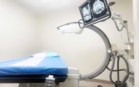
Fluoroscopy is X-ray imaging shown as a moving picture rather than as a single photograph. The C-arm is the machine that produces it — a C-shaped frame carrying the X-ray tube at one end and the detector at the other, positioned around the patient so that bone, needle and contrast are visible on a screen while the needle is advanced. It is not a diagnostic scan and it is not read like one. Its purpose is to put something exactly where it was meant to go.
Why guidance is used
-
Surface landmarks are not reliable enough
Studies of injections placed by feel alone find that the needle frequently ends up somewhere other than intended, including in experienced hands. The usual consequence is not harm — it is a procedure that did not work, and a false conclusion drawn about the structure that was supposedly treated.
-
Contrast confirms where the medication will go
A small volume of radiographic contrast is injected before the medication. Its spread on the screen shows whether the intended structure will be reached, and whether the needle tip has entered a blood vessel — which would otherwise send the injection somewhere it should not be.
-
The level has to be right
In the spine, adjacent levels are a centimetre apart and produce different symptoms. Imaging is what establishes that the needle is at the level the examination implicated.
-
Depth
The targets are deep, and several of them sit close to structures that must not be entered. Seeing the needle in relation to bone is what keeps the margin.
Procedures it guides
-
Epidural steroid injection
Interlaminar, transforaminal and caudal approaches are all performed under fluoroscopy, with contrast confirming epidural spread.
-
Medial branch block and facet joint injection
The targets are small bony landmarks on the vertebra. These are difficult to reach reproducibly without imaging, and reproducibility is the entire value of a diagnostic block.
-
Selective nerve root block
Placing medication at one specific nerve root, both to treat it and to establish whether that root is the source of the pain.
-
Sacroiliac joint injection
The joint surface is oblique and irregular, and blind injection into it is unreliable.
Radiation
A fair question, and one worth answering with numbers rather than reassurance.
-
How much
A guided spinal injection uses brief, intermittent imaging rather than continuous exposure. The dose for a routine procedure is of the same order as a small number of plain X-rays of the same region.
-
How it is kept down
Pulsed rather than continuous screening, collimation to the area of interest, keeping screening times short, and using stored images rather than live ones where a still picture will answer the question.
-
Pregnancy
Fluoroscopy is avoided in pregnancy. Any possibility of it should be raised before the procedure, not during it.
What it cannot do
-
It does not show soft tissue
Discs, nerves, tendons and muscle are effectively invisible on fluoroscopy. Questions about them are answered by MRI or by ultrasound, and guidance for soft-tissue targets is usually ultrasound for the same reason.
-
It is not a diagnostic study
A fluoroscopic image is a working picture taken to steer a needle. It does not replace imaging performed and reported to answer a diagnostic question.
-
Contrast has its own considerations
Iodinated contrast is used in small amounts. A previous reaction to contrast, or significantly impaired kidney function, changes what is appropriate and should be known beforehand.
Common questions
Is the radiation dangerous?
The dose from a routine guided injection is small and is weighed against performing the same procedure without knowing where the needle is. Repeated procedures over a short period are worth discussing, and dose is recorded.
Am I asleep for it?
No. These procedures are done awake with local anaesthetic, partly because your response during the injection is useful information.
Does guidance make it hurt less?
Not directly. It makes the injection accurate, which is a different thing — though a needle that reaches its target on the first attempt generally involves less probing on the way.
Could ultrasound be used instead?
For soft-tissue and superficial joint targets, often yes, and it avoids radiation entirely. For deep spinal targets where bone is the landmark and vascular spread has to be excluded, fluoroscopy remains the standard.
General reference information, and not advice about your own case. It may not reflect the most recent guidance or what is offered on a given day — call (201) 408-5151 to check.
Schedule an appointment
Dr. Yoo Rehab Clinic welcomes new patients from across Northern New Jersey and New York City, including Englewood, Teaneck, Fort Lee, Palisades Park and Ridgefield. Call today to learn more about insurance, scheduling, and how we can help.